
DEVELOPMENT OF VARIOUS OCULAR STRUCTURES
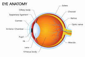
Vitreous
Primary or primitive vitreous. Itemonymil in orgn and icx vocale structure heong the bryalid systems of the vessels
Cornea
pablomdowed from du fic ecudem. Dakhelm Descemet’s brine, strom and Secondary or definitive or vitreous proper. It is wered by the news toderm of the optic cup Bean’s layer are derived from the sul crest Tertiary vitreous It developed from the neuroectoderm in the ciliary region and is represented by the zomulic
Sclera
Develes from the malcrest rescichyme that surrounds the optie cop
Eyelids
Develop by the reduplication of the surface ectoderm above and below the cornea
Choroid
Derived from die inner vascular layer of the endyme Conjunctiva Develop from the resoderm lang the lids and covering that surrounds the optic cit the globe
Ciliary Body
The new layers of the epiti id the cillary body develop front thac anterior parte o layer of th opoc cup (nciroccoden Stronts of the olaty body cility uncle and blood
Lacrimal Apparatus
Lacrimal gland It is derived from the epithelial hade which grow from superolateral side of conjunctival sac Lacrimal sac, sasolacrimal duct and canaliculi They develop from the surface ectoderm venchs are derived from the ular teyer of the mesenchymne
Iris
All extraocular muscles develop from myotomic celle of the Both epithelial layers develop from the martial region per-optic meseslerinal-samites that have shifted cranially of the optic cup (neurocctodermo)
• Sphincter and the dilator pupillae muscles are derived from the anterior epithelium (neurocioderm)
The Features of the Eye at Birth Stroma and blood vessels develop from the vascular imesenchyme present anterior to the cup
The sycball is small, the anteropostetice dieter (asal
length) is about 16 mm, and it is fully developed at the age of 6 ycars
Retina
- Corneal diameter is 10 mm Anterior chamber is shallows
- It develops from two walls of optic cup Sensory renna from the inner wall.
- The newborn is hypermetrope by 230 diopter.
- Retinal pigment epithelium from the outer wall.
- The pupil is small and does not dilate fully