
Applied Anatomy and Physiology of the Eye
GROSS ANATOMY
The anomy and physiology of the individual structure of the eye is described in detail at the beginning of cachi chapter. Here, the gross anatomy is described The structure of the eye includes
• Orbital Cavity-protects the eyeball Eyeball-organ of vision
Extraocular muscles-move the eyeball in different directions of gaze Fascia and fat-provides support to the eyeball
EYEBALL
Eyeball is the organ of vision lying in the orbital cavity which protects it from the external injuries. The eyeball coasts of three layers or coats
Corneoscleral (Fibrous) Layer
It gives shape and protection to the structures of eyeball. It is divided into the following parts Com A transparent structure that comprises anterior one-sxth of the outer layer.
Selera: A dense, fibrous, collagenoirs structure that fluid for the transmission of light rays comprises posterior five-sixth of the outer layer
uLimbur A transitional zone about 2 mm wide that
Uveal (Vascular) Layer It is divided into three parts:
Cervid: A vascular layer, which provides nutrition to the outer layers of retina.
Ciliary body Produces aqueous humour through its olury processes and takes part in accommodation through the muscular portion. m
Acts as a diaphragm with a central aperture called pl, which controls the amount of light entering the eye.
Retina (Nervous) Layer
In this nervous tistic layer te retina, the light energy w converted into the electrical energy (action potcunal) which is transmitted via the visual pathway to the occipital
cortex. It consists of
L Sensory rema . Retinal pigment epithelium
The Interior of the Eyeball It is divided into two segments
Anterior Segment
It is further divided by iris diaphragm unto two chambers
Anterior Chamber
It is a small cavity, which is bounded anteriorly by the
cornca, posteriorly by the front surface of iris and lens, peripherally by the anterior chamber angle. The anterior chamber depth in central portion is about
2 to 3 mm, its volume is about 0.25 ml and contains a clear fluid called aqueous humour
It provides nutrition to the vascular structures such as lens, trabecular meshwork and cornea. It is transparent
ii. Posterior Chamber
contains trabecular meshwork through which aqueous. It is small slit like cavity-bounded anteriorly by the ires, posteriorly by the anterior surface of the lens and zonules and penpherally by ciliary process. It contains about 0.06 ml of aqueous humour. Imour is drained.
Posterior Segment (Vitreous Cavity)
It is bounded anteriorly by the lens, zonules and caliary process and posteriorly by retina and optic nerve.
Its volume is about 4,5 ml and contains the vitreous humour, transparent jelly like structure composed of 99% water and 1% solds.
. It is transparent for the passage of light ray
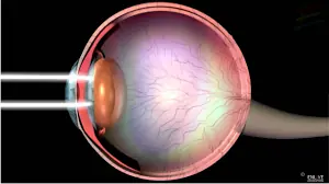
21 Structure of the eye ball
Optic nerve
Crystalline Lens
It is a biconvex grossly transparent structure located directly behind the mes and pupil and in front of the vitreous body. It takes part in the refraction of light rays and accommodation.
Extraocular Muscles
They are ses in number, four recti and two oblique muscles, which route the cychall in different directions of gaze that so one can see in a wide range without turning the face.
Fat and Fascia
It provides mechanical support to the eyeball and suspends in the center of the orbit.
Eyelids
Each upper eyelid consists of tarsal plates, Muller’s muscle, leator palpebrac superioris and various glands mainly, the Meibomian glands and the glands of Zeis and Moll. It gives mechanical protection from the external injuries and its glands produce the aqueous and lipid layers of the tear film.
Conjunctiva
Itna thin translucent membrane, which joins the eyeball with eyelids. It contains goblet cells and accessory lacrimal glands. Their secretions contribute to mucin and aqueous layer of tear film
Tear Film
The precorneal tear film is composed of three layers: Superficial lipad, middle aqueous and inner micin layer It converts irregular corneal surface into smooth optical surface, and prevents the drying up of corneal surface.
Lacrimal System
Lacrimal system consists of Aqueous producmg glands, the main lacrimal gland lying in the lateral part of the roof of the orbit and the accessory lacrimal dlands present in the superior formix of the conjunctiva. Drainage system that consists of Puneti, canaliculi, common canaliculus Escrimal ste, and the nasolacrimal duct, which opens into the inferior meatus of the nose.
- It drains the rear film
- Optic Nerve and Visual Pathway
- Optic nerve is formed by the axons of the ganglion cells and arises from the back of the eyeball The two optic nerves join together to form the optic chiasma, where nasal fibers of each half of the retina decussate. The optic tract arises from the optic chiasma, carries the ipsilateral temporal fibers and the contralateral nasal fibers of each retina that terminates in the lateral geniculate body.
- The optic radiation arises from the lateral gemculate body and terminates in the visual occipital cortex, which is located on the medial aspect of the occipital lobe above and below the calcarine sulcus.
Pupillary Reflex Pathway
The nerve fibers arise from the retina; travel in the optic nerve, chiasma and optic tract to enter into the pretectal nucleus. These fibers do not terminate in the lateral geniculate body.
The pretectal nucleus fibers project to both, the ipsilateral and the contralateral Edinger-Westphal nucleus, which is the parasympathetic nucleus of the oculomotor nerve.
From Edinger Westphal nucleus, they travel in the oculomotor nerve and synapse in the ciliary ganglion. The postganglionic nerve fibers from ciliary ganglion travel via the short ciliary nerves to supply the sphincter pupillac muscle.
Motor Nerve Supply
Oculomotor (thurd) nerve supplies all the extraocular muscles (except superior oblique and lateral rectus), levator palpebrae superioris and smooth muscles concerned with accommodation namely sphincter papillac and ciliary muscles
Trochlear (fourth) nerve supplies the superior oblique muscle.
• Abducent (sixth) nerve supplies the lateral rectus muscle.
Sensory Nerve Supply
The ophthalmic division of trigeminal nerve through its various branches supplies all structures of the eyeball and adnexa.
Blood Supply
The ophthalmic artery through its various branches supplies the extraocular muscles, the structures of eyeball, and adnexa.
Tags: Anatomy and Physiology of the Eye, anatomy of eye, structure of eye